


Introducing our revolutionary CT technology, CLARIS V. This state-of-the-art cone-beam CT system is meticulously crafted to prioritize patient safety by minimizing x-ray exposure while delivering exceptional image quality. With CLARIS V, you gain access to a low-dose CT machine that combines user-friendly operation, streamlined patient throughput, and advanced cardiac imaging capabilities, all complemented by a wide range of post-processing and diagnostic features.
Each CLARIS V system includes an integrated workstation equipped with the powerful XC acquisition and Clarity Viewers software suite. This comprehensive package empowers you with robust diagnostic tools, enhancing patient care through image acquisition, annotation, and manipulation capabilities.
17" x 17"
Field of View
16 Bit
Flat Panel Detector
430 mm x 430 mm
Sensor
140 μm
Voxel Size


Finally an Equine Trailer with full Accessibility:
The E-Claris V Mobile Units for Equine can reach remote areas, u, disaster-stricken regions, or events where immediate equine imaging is necessary.

CLARIS V | Real Use Images


CLARIS V | Use Cases
The Claris V is designed specifically for veterinary use are not only available they are becoming increasingly common in veterinary practices and clinics.

ORTHOPEDICS
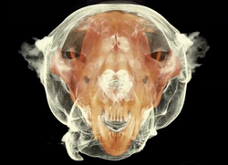
Claris V can be used to assess musculoskeletal conditions in animals, including fractures, joint diseases, and skeletal deformities. It provides detailed images of bones and joints, enabling veterinarians to diagnose and plan treatments, such as orthopedic surgeries or the fitting of orthopedic devices.
SOFT TISSUE IMAGING
Claris V can also be utilized for imaging soft tissues in animals. It can assist in evaluating masses, tumors, or abnormalities in organs or soft tissue structures.
NASAL AND SINUS EVALUATIONS
Claris V is valuable for examining the nasal and sinus cavities in animals. It helps in identifying nasal masses, foreign bodies, or structural abnormalities that may be causing respiratory issues or other related conditions.
PRE-OPERATIVE PLANNING
Claris V imaging allows veterinarians to perform detailed pre-operative planning for complex surgeries. It provides precise 3D visualization of the anatomical structures involved, aiding in surgical decision-making and reducing the risk of complications during procedures.
DENTAL & ORAL EXAMINATIONS
Claris V is particularly useful for evaluating dental and oral conditions in animals, such as tooth root infections, dental fractures, and oral tumors. It provides detailed 3D images of the oral cavity, allowing veterinarians to plan and perform dental procedures more accurately.
CLARIS V | Generate Revenue
Adding CLARIS V (CBCT) machine to a veterinary practice can be a valuable investment and offer various revenue-generating opportunities.

$1,050
Average Charge
Per Scan
The 3DCT imaging modality provides diagnostic clarity that transforms your treatment capabilities.
Monthly
Easy Financing &
Monthly Payments
15 mos.
Claris V
Pays For Itself
Claris V will provide enough data in a single scan to reduce multiple anesthesia and ease stress on the patient and owner.
Claris V is a revenue producer. Learn how clinics are transforming patient care while the Claris V pays for their practice.
Finally, a compact, and affordable CT, that's easy to install and
is a revenue producer for your practice.
DIAGNOSTIC IMAGING SERVICES
CLARIS V providing detailed three-dimensional scans for enhanced diagnostic capabilities. Charging for CBCT scans and interpretations can help diagnose complex dental, oral, cranial, and skeletal conditions accurately.
REFERRAL CENTER
CLARIS V become a referral center for veterinarians without access to CBCT technology. By offering CBCT services to their clients, you can attract referrals and charge a fee for conducting scans and providing reports.
3D PRINTING & ADDITIONAL SERVICES
CLARIS V can serve as an additional revenue stream by offering other related services. For example, you can provide 3D printing services to create patient-specific anatomical models based on CBCT scans, which can be used for surgical planning or client education. Charging for such value-added services can contribute to revenue generation.
CLARIS V, the revolutionary all-in-one cone-beam CT system transforming diagnostic imaging. With advanced Cesium detector technology, it combines the functionalities of a traditional x-ray room, fluoroscopy, and CT imaging. Offering streamlined workflow, it enables various studies such as chest, cranial, dental, and orthopedics in a single solution.
CLARIS V's 17" by 17" field-of-view, powered by Cesium Sensor Technology, delivers exceptional chest images for early cancer detection. Experience the ultimate in veterinary care with CLARIS V's comprehensive cone-beam CT capabilities.

Larger, Consistent Field-of-View
Traditional flat panel detectors have a limited field-of-view and can miss vital anatomical information, the Claris V captures more important diagnostic image data in a single exposure with its large format 17X17 inch sensor. Cesium Sensor technology reduces dose and improves CT resolution at very high frame capture rates, reducing movement artifacts and improving patient outcomes.
✔
✔
Emergency Care, Surgical Planning, General Imaging Large 17” by 17"
Detector Plates, Minimal Footprint, Ideal For Small Clinics, Dose Optimized By Imaging Application.
✔
✔
✔
Variable Resolution Down To 140-micron Voxel Size.
Advanced CBCT Capable Of Full Chest Studies. Traditional Full CT, Tomosynthesis.
Replace X-Ray Rooms


Axis CT Exam Table
The New Axis Motorized Table Options Allow:
-
Motorized Z Axis Adjustment
-
Robotic X Movement
-
Mobile
-
Attaches to CT Gantry with Locking Interface
-
Radio Translucent
-
Low Attenuation Carbon Fiber Table Top
-
Manual or Robotic Movement
-
Adjustable Height

 |  |  |
|---|---|---|
 |  |  |
 |  |  |
 |  |